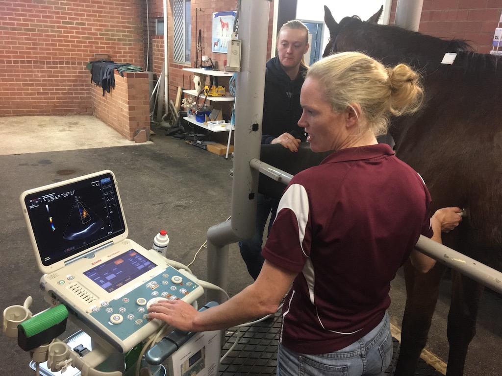
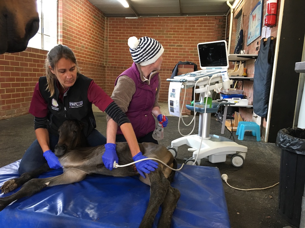
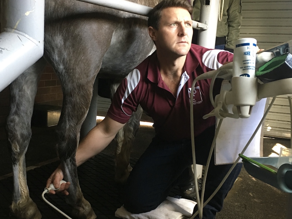
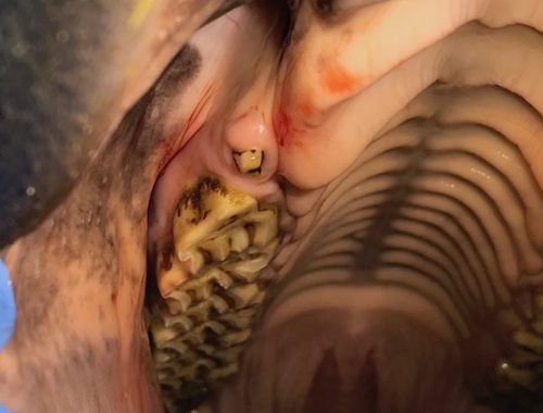
Ultrasound is a highly valuable, non-invasive, diagnostic tool that plays a vital role in equine veterinary care. At Ascot Equine Veterinarians, our skilled team utilises advanced ultrasound technology, including multiple machines and a variety of transducers, to examine a wide range of body systems in horses.
Ultrasound in Lameness Diagnosis and Monitoring
Ultrasound is used widely in lameness examinations to evaluate soft tissue structures of the limbs, such as tendons, muscles and ligaments. This method allows us to detect injuries and monitor the healing process, guiding effective treatment. Ultrasound also provides detailed images of bone and joint surfaces, helping to confirm fractures in areas that are difficult to assess with radiographs, such as the pelvis, ribs and scapula. Additionally, we use it to evaluate wounds, soft tissue swellings, and other abnormalities.
Abdominal Ultrasound
Abdominal ultrasound is used routinely in the workup and assessment of colic cases. It is also an important diagnostic tool in the investigation of horses with unexplained weight loss and can assist in the detection of tumours, cysts and abscesses. When deeper investigation is needed, we can use ultrasound to safely guide biopsies of internal organs, such as the liver, lungs, and kidneys.
Cardiac Ultrasound
Ultrasonography of the heart, know as echocardiography, allows us to evaluate the heart’s chambers and valves. This is particularly important for assessing the significance of heart murmurs. Colour flow Doppler is used to assess dynamic blood flow through different parts of the heart in real time, enabling early detection of issues like valve dysfunction. Ultrasound is also valuable in assessing peripheral blood vessels and detecting blood clots (thrombi).
Thoracic Ultrasound
Thoracic ultrasonography plays a critical role in assessment of the thoracic cavity, including the lungs, is important in the assessment of horses with pneumonia, lung masses or pleural effusions. Early signs of fluid buildup or surface changes in the lungs can be identified, allowing for timely and effective treatment.


